Always refer to the main Pearson general guidelines and science guidance when writing alternative text for images.
When describing Anatomy & Physiology images, keep in mind the following key aspects and examples. Not all guidance will apply to all images.
- Identify and describe the different structures or parts of the image using the terminology from the main text. This includes naming the organs, tissues, cells, and other structures that are visible in the image.
- Explain the function of each structure. This includes describing how the structures work together to carry out specific physiological processes, such as digestion, circulation, and respiration.
- You may need to provide context by explaining where the structures are located in the body and how they relate to other structures and systems.
- Only include relevant details, such as the size and shape of the structure. These details can help clarify the image and provide understanding.
Examples
Reminder: The Mastering authoring platform has a title field and alt text field but does not have the functionality for a long description. Alternative text descriptions in Mastering may have more than 255 characters. The eText 2 authoring platform has the functionality for alt text and a long description. For more information about the different authoring systems, see Platform Authoring Information.
Example 1
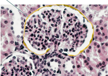
Alt Text
A cross-sectional histological view of a tissue containing a mushroom-shaped structure, separated by white space from the rest of the tissue. The structure is composed of pink elements, each with a very dark large blob inside it. The outer border of the white gap is highlighted, and resembles an extremely thin layer of highly flattened elements.
Example 2
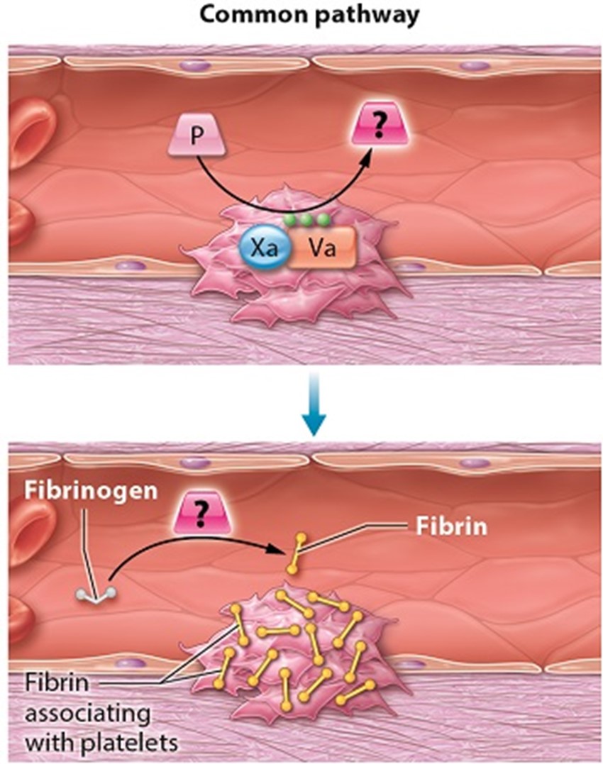
Alt Text
A diagram illustrates the common pathway of coagulation. Factor 10 A and factor 5 A form a complex at the platelet plug. This complex then acts on prothrombin to produce an unknown enzyme. The unknown enzyme converts fibrinogen into fibrin, which associates with the platelets in the plug.
Example 3

Alt Text
Posterior view of the upper arm. The highlighted head of the triceps brachii muscle is in the middle of the upper arm.
Note: The image was for an assessment question: Identify the highlighted head of the triceps brachii muscle.
Example 4
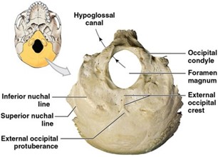
Alt Text
An inferior view of the occipital bone.
Long Description
(Must be marked up in HTML)
The occipital bone forms most of the posterior skull base. The inferior view shows 7 occipital bone structures. The following table identifies whether each structure is bilateral. The table also provides a description of each structure’s shape and location.
| Structure | Bilateral | Description |
|---|---|---|
| Foramen magnum | No | An oval opening in the medial anterior occipital bone |
| Occipital condyle | Yes | A rounded protuberance on the lateral anterior edge of the foramen magnum. |
| Hypoglossal canal | Yes | A foramen that extends medially through the lateral anterior edge of the foramen magnum, superior to the occipital condyle. |
| External occipital crest | No | A midline ridge posterior to the foramen magnum. |
| External occipital protuberance | No | A V shaped structure at the posterior end of the external occipital crest. The V opens posteriorly. |
| Inferior nuchal line | Yes | A curved ridge roughly parallel to the edge of the foramen magnum. The ridge extends from the anterior margin of the occipital bone to a point lateral to the external occipital protuberance. |
| Inferior nuchal line | Yes | A curved ridge along the margin of the occipital bone, lateral to the inferior nuchal line. |
Example 5
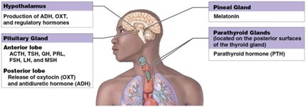
Alt Text
A diagram shows the hormones secreted by glands in the endocrine system.
Long Description
(Must be marked up in HTML)
The following table provides the location and hormones for each gland shown in the diagram. Except for the posterior lobe of the pituitary, all glands produce the hormones they release.
| Gland | Location | Hormones |
|---|---|---|
| Hypothalmus | Inferior to the thalmus and superior to the brainstem | Antidiuretic hormone (A D H) Oxytocin (O X T) Regulatory hormones |
| Pituitary gland, anterior lobe | Inferior to the hypothalmus and anterior to the brainstem | Adrenocorticotropic hormone (A C T H) Thyroid stimulating hormone (T S H) Growth hormone (G H) Prolactin (P R L) Follicle stimulating hormone (F S H) Luteinizing hormone (L H) Melanocyte stimulating hormone (M S H) |
| Pituitary gland, posterior lobe | Inferior to the hypothalmus and anterior to the brainstem | Releases oxytocin (O X T) and antidiuretic hormone (A D H) |
| Pineal gland | Posterior to the third cerebral ventricle in the midline | Melatonin |
| Parathyroid glands | Posterior surface of the thyroid gland in the neck | Parathyroid hormone (P T H) |
Header text
Example 6
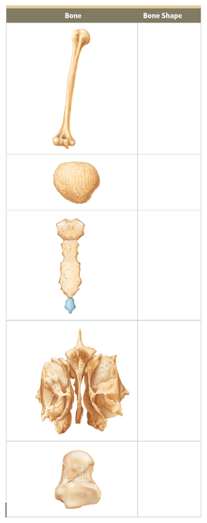
Alt Text
A table for identifying bone shapes.
Long Description
(Must be marked up in HTML)
The table has 6 rows and 2 columns. Row 1 contains the following column headers. Column 1, bone. Column 2, bone shape. Column 1 contains illustrations of different bones. Column 2 is blank. The bones in column 1 are as follows.
- Row 2. A long bone with a ball-shaped tip and an inverted heart-shaped tip at either end.
- Row 3. A smooth oval bone.
- Row 4. A long, flat, necktie-shaped bone..
- Row 5. An irregular-shaped bone with layers and sharp tips
- Row 6. A small flat bone with round protuberances at the edges.
Example 7
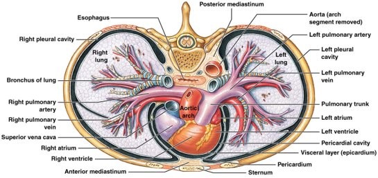
Alt Text
A superior view of the thoracic cavity shows structures of the cardiovascular and respiratory systems.
Long Description
(Must be marked up in HTML)
In the superior view, the thorax is cut along a plane above the aortic arch, and the anterior side of the body is on the bottom.
The heart is at the medial anterior of the thorax, separated from the sternum by the anterior mediastinum. The outermost, visceral layer of the heart is the epicardium. The pericardial cavity separates the epicardium from the pericardium, which forms the outer wall of the cavity. The heart is positioned so that the ventricles are partially anterior to the atria and the right atrium is partially anterior to the left atrium. From right to left in the anatomical reference frame, the superior vena cava, aortic arch, and pulmonary trunk rise from the posterior side of the heart.
The pulmonary arteries extend laterally from the superior end of the pulmonary trunk. Along with the pulmonary veins and bronchi, the arteries form overlapping treelike structures that branch into the lungs’ spongy tissue. The lungs are located inside the pleural cavity.
As its name suggests, the posterior mediastinum is located posterior to the heart and medial to the lungs. Within it, the esophagus appears as an oval compressed along an anterior to posterior axis. The esophagus is immediately anterior to the spinal column and medial to the aorta.
Example 8
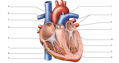
Alt Text
Structure of the heart.
Long Description
(Must be marked up in HTML)
A diagram of the heart with blank labels.
- Part A. Large oxygenated vessel that connects to the left atrium.
- Part B. The valve that empties blood out of the right atrium.
- Part C. The large deoxygenated vessel that brings blood to the right atrium from above.
- Part D. The top right chamber of the heart.
- Part E. The valve between the right atrium and the right ventricle.
- Part F. Bottom right chamber of the heart.
- Part G. Muscles within the walls of the ventricles that pull open the valves.
- Part H. The large deoxygenated vessel that brings blood to the right atrium from below.
- Part I. Large deoxygenated vessel that brings blood to the lungs from the right ventricle.
- Part J. Vessels that bring blood into the heart from the lungs.
- Part K. Top left chamber of the heart.
- Part L. Valve that opens to empty blood from the left ventricle.
- Part M. Valve that empties blood from the left atrium into the left ventricle.
- Part N. Stringlike structures that connect a valve to the ventricle wall.
- Part O. Bottom left chamber of the heart.
- Part P. Wall between the two ventricles.
Dated: 2023-12-01
